
Seeing the Invisible
The Genius of Phase Contrast Microscopy | Part 8/30

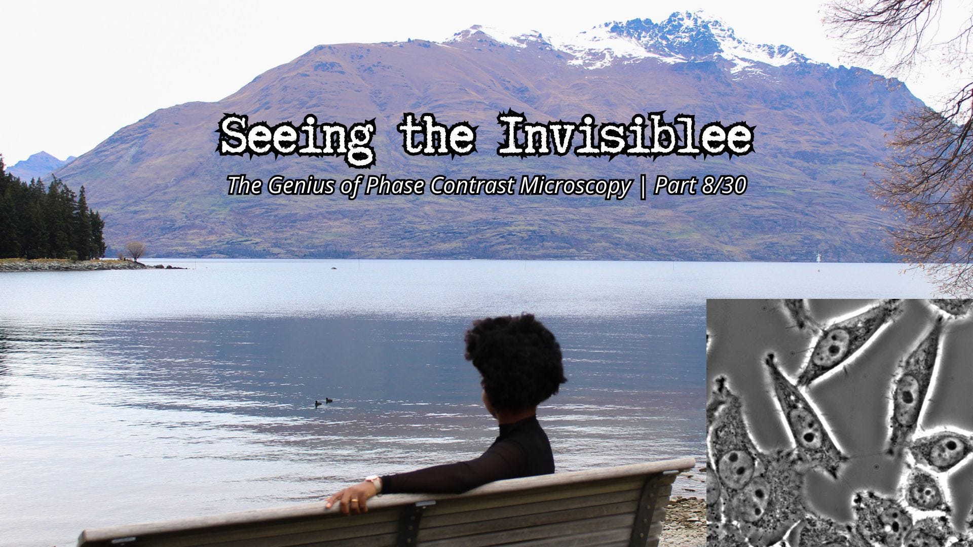
A picture of me at Queenstown Research Week, New Zealand and a phase contrast microscope image of a cell. Credit: Science Equipment
After over one thousand applications to so many countries and universities and 3 years later, I got my yes, actually, three yeses. One for a fully funded PhD scholarship to New Zealand, generously covering tuition and living expenses for 3 years and funded by the Doctoral Office. We are talking of over $NZ 120,000 worth of funding. A massive thank you to The University of Otago! The other scholarship was for a fully-funded scholarship to Japan under the JICA scholarship scheme. The third was also a fully-funded scholarship by The Department of Physiology, University of Otago, New Zealand (forfeited for the Doctoral Office’s scholarship). I considered both options and went for the New Zealand option. Sometimes, I consider this sojourn as a training ground, an isolating 4 years needed for my strengthening, pruning and grounding.
The strength of character developed in New Zealand laid a solid foundation for my becoming a better mum, thorough scientist, professional and fundamentally, extremely empathetic. Positively and unashamedly biased towards my fellow women. We have it tougher and we would cover more grounds if we worked together.
My good friend of over 19 years, Dr Chidozie Ojobor Gut Health Simplified, who has now co-built a multi-million dollar company in Canada, Vitract, has been ringing it in my ears for over 10 years now to write a book. That, I have now started doing. As we mark your birthday Dozie, I use this opportunity to say a big thank you. Thank you for seeing a story when all I saw at that point in time was severe discomfort. Y’all, you need a Dozie in your corner.
I recently interviewed Chidozie on how he built his multi-million dollar company (backed by New York TechStars some years ago). The video will be up on my YouTube channel in roughly 2 weeks’ time.
Welcome to Week 8! Today, we have now officially crossed the threshold into Phase 2: Illuminating Biology & Breaking the Limit.
Over the past seven weeks, we mastered the basic physics of light and even built our classic wide-field microscope in Part 7. Studying the intricate mechanics of cardiovascular physiology, the standard wide-field microscope presented a massive, frustrating hurdle to me still.
I always did not fancy looking at dead, chemically preserved biology tissues; I also wanted to look at living cells. How about seeing life in motion. But if you take a living, unstained cell and place it under a standard light microscope, do you know what you see?
Not much details. A living unstained cell under a standard light microscope appears faint and ghost-like. Most of its internal structures are nearly transparent to visible light and differ only slightly in refractive index from the surrounding cytoplasm, so very little contrast is produced.
It is like trying to find a clear ice cube in a glass of water. Because cells are mostly made of water, they are almost completely transparent. For a long time, the only way biologists could see cellular structures was to kill the cell (fixation) and dye it with harsh, colourful chemical stains. But dead cells do not beat nor contract like living heart cells for instance. To truly push the borders of physics with biology as an igniting fuel, we needed a way to see the invisible without necessarily destroying it.
The Microscopic Speed Bump (The Core Concept)
In the 1930s, a Dutch physicist named Frits Zernike came up with a brilliantly elegant solution that revolutionised biology. Zernike knew that while a transparent cell doesn’t absorb light (which is why it doesn’t cast a shadow), its internal structures, like the nucleus or the dense cell membrane, are slightly thicker and denser than the water surrounding them.
When a wave of light travels through that denser cellular material, it hits a microscopic speed bump. The light slows down just a tiny fraction of a second compared to the light travelling only through the surrounding water. In physics, this tiny delay is called a phase shift.
The catch: human eyes and camera sensors are completely blind to phase shifts. We can only see changes in colour (Ultraviolet to red wavelength a.k.a rainbow) or brightness (amplitude). We cannot see “time delays” in light waves. We simply cannot.
Zernike’s Optical Magic
Zernike’s genius was finding a way to convert an invisible phase delay into a visible difference in brightness. When light passes through a living cell, some light waves slow down slightly because different parts of the cell have different refractive indices and thicknesses. Although these waves become delayed in phase, the delay is normally invisible to the human eye because our eyes detect brightness, not phase shifts.
To solve this, Frits Zernike placed a specially engineered optical element called a phase ring inside the microscope. The phase ring shifted the phase of the undeviated background light so that it could strongly interfere with the diffracted light emerging from the specimen.
Once these waves recombined, tiny phase differences were transformed into visible intensity differences through interference. In some regions the waves reinforced each other (constructive interference), while in others they partially cancelled out (destructive interference). Suddenly, structures that had been nearly invisible appeared with dramatic contrast.
Now, through the eyepiece, the transparent living cell emerged with dark shadows, bright details, and the characteristic glowing halos of phase-contrast microscopy outlining its structures and organelles.
This was an extraordinary breakthrough in optical physics and biological imaging, a feat that earned Zernike the Nobel Prize in Physics in 1953.
Why It Matters: Watching Life Unfold
This was a remarkable mindset shift for the biological sciences. With Phase Contrast microscopy, time-lapse imaging was now a reality and time did not have to be frozen to capture life’s intricate details.
We no longer had to act as cellular morticians, killing (fixing) and staining our samples just to see them. We could place a petri dish of cardiovascular cells under the lens and watch them contract and beat in real-time. We could watch a single cell divide into two (mitosis) and we could excitedly watch immune cells actively hunt down, engulf, and digest bacteria, a process known as phagocytosis.
Phase contrast gave us a non-invasive, non-intrusive window into the living, breathing mechanics of the cellular universe. It is a perfect example of how an intricate understanding of physics can break open an entirely new frontier in biology.
Next Week...
But while Phase Contrast is incredible for showing us the overall architectural blueprint of a living cell, it has a limitation. It does show us the walls, the doors, and the rooms, but it doesn’t tell us who is inside those rooms. We love to pokenose, remember? That’s all scientists do, ask questions, poke, probe, prove…
What if we don’t want to see the whole cell? What if we want to track one specific, tiny cardiovascular protein as it moves through that living architecture?
So, next week, we are entering my absolute happy place. I guess you can tell already. In Week 9 titled, Lighting Up the Cell, we are going to dive into the vibrant, glowing world of Fluorescence Microscopy.
If you had a Phase Contrast microscope on your desk right now, what living thing would you want to look at first? Let me know in the comments!
Relevant Links
Phase 1: The Physics of Light & The Genesis of Sight
Part 1/30: The Catalyst: From a Biology Basement in New Zealand to UK Biophysics
Part 2/30: The Genesis of Sight: How We First Peered into the Cellular Universe
Part 3/30: The Physics of Light: Why We Use Photons as a Measuring Stick
Part 4/30: Bending Light to My Will: The Magic of Lenses and Mirrors
Part 5/30: The Secret Sauce of Clarity: Understanding Numerical Aperture
Part 6/30: The Physical Barrier of Light: Breaking Down the Abbe Diffraction Limit
Part 7/30: The Classic Workhorse: Inside the Wide-Field Microscope
Dr Chidinma Okolo is a manager at a National Space organisation, an accomplished beamline scientist at a National Science Facility, and the lead creative behind “The Quiet Learner” brand. A cardiovascular physiologist by doctoral training, a physicist by synchrotron experience, and a business/project strategist by apprenticeship/hands-on training. A typical example of a polymath. All skills jointly enabling her to sit as a project delivery professional and a subject matter expert at the intersection of biology and microgravity; seamlessly bridging the divide between biology, physics, business, projects and the end consumer. In her current cross-cutting role, she sits at the triple helix intersection between academia, industry and government.
Chidinma is passionate about promoting women in STEM as well as black African and minority ethnic immigrants and ensuring that both minority, under-served and underrepresented groups can move jobs across sectors when they need to, without feeling stuck. She strives to achieve this by equipping them with appropriate tools in the form of lifelong learning, visibility, sponsorship/mentorship and intentional networking strategies to amplify their voices and brands. Chidinma uses emotive and simplified storytelling to carry her readers, viewers and audience along. Providing proven frameworks, tested models and insights to define their own career, entrepreneurial and creative paths. You can check out her video blogs, The Quiet Learner and The Career Lab with Dr Chidi on YouTube. Chidinma writes in her own capacity and her opinions are solely her own.
© Dr Chidinma Okolo
2026